Tendon Diagram : Achilles Tendon Rupture And Achilles Tendonitis Treatment : Intermediate back muscles and c.. Allows the action of raising the foot. The hand incorporates countless muscles, bones, tendons and ligaments into simple motion and this chart covers them all. Lower back muscle diagram anatomy does degenerative disc disease affect the lower back muscle? Bones, muscles, tendons and nerves which will each give slightly different foot pain symptoms. Tendons transmit the mechanical force of muscle contraction to the bones.
Learn about the anatomy and physiology of tendons. May 11, 2021 · muscles of lower back diagram in this image, you will find an occipital bone, sternocleidomastoid, trapezius, deltoid in muscles of the lower back diagram. Allows the foot to be turned inward and also supports the arch of the foot. The achilles tendon is also called the calcaneal tendon. What are some examples of tendons?
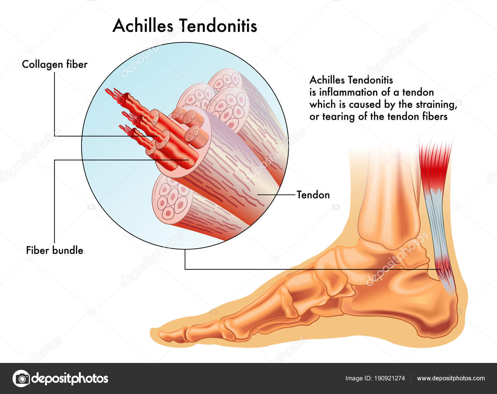
Achilles Tendon Wikipedia from upload.wikimedia.org Bones, muscles, tendons and nerves which will each give slightly different foot pain symptoms. Also allows the action of raising up onto toes. Foot anatomy diagram, foot joint diagram, foot sprain diagram, foot tendons and ligaments pain, leg tendon diagram, peroneal tendonitis, foot, foot anatomy diagram, foot joint diagram, foot sprain diagram, foot tendons and ligaments pain, leg tendon diagram, peroneal tendonitis. More images for tendon diagram » A foot pain diagram is a great tool to help you work out what is causing your ankle and foot pain. Allows the foot to be turned inward and also supports the arch of the foot. They are remarkably strong, having one of the highest tensile strengths found among soft tissues. Its muscle belly is in the forearm.
It attaches to the wrist bone, the pisiform, and as well as the 5th hand bone.
Bones, muscles, tendons and nerves which will each give slightly different foot pain symptoms. Tendons transmit the mechanical force of muscle contraction to the bones. Jul 12, 2020 · muscle anatomy for gym 12 photos of the muscle anatomy for gym muscle anatomy and fitness, muscle anatomy for fitness, muscle anatomy for gym, human muscles, muscle anatomy and fitness, muscle anatomy for fitness, muscle anatomy for gym. Learn about the anatomy and physiology of tendons. Its muscle belly is in the forearm. The tendon travels along the inside of the forearm on the side of the small finger and crosses the wrist. What are the major tendons in the body? Allows the foot to be turned inward and also supports the arch of the foot. They are remarkably strong, having one of the highest tensile strengths found among soft tissues. Allows the action of raising the foot. The hand incorporates countless muscles, bones, tendons and ligaments into simple motion and this chart covers them all. Attaches the calf muscles to the calcaneus, most important muscles for running, jumping, walking etc. There are a whole range of structures e.g.
Lumbar sprain occurs when ligaments are overstretched or torn. The tendon travels along the inside of the forearm on the side of the small finger and crosses the wrist. There are a whole range of structures e.g. Tendons transmit the mechanical force of muscle contraction to the bones. The fcu tendon is one of two tendons that bend the wrist.
Vector Illustration Achilles Tendon Image Foot Anatomy All Tendons Bones Vector Image By C Rob3000 Vector Stock 190921274 from st3.depositphotos.com They are remarkably strong, having one of the highest tensile strengths found among soft tissues. The hand incorporates countless muscles, bones, tendons and ligaments into simple motion and this chart covers them all. How many tendons are in the body? Learn about the anatomy and physiology of tendons. It attaches to the wrist bone, the pisiform, and as well as the 5th hand bone. More images for tendon diagram » A foot pain diagram is a great tool to help you work out what is causing your ankle and foot pain. Its muscle belly is in the forearm.
Learn about the anatomy and physiology of tendons.
Allows the action of raising the foot. The tendon travels along the inside of the forearm on the side of the small finger and crosses the wrist. Chloe wilson bsc(hons) physiotherapy reviewed by: There are a whole range of structures e.g. Lumbar sprain occurs when ligaments are overstretched or torn. How many tendons are in the body? Attaches the calf muscles to the calcaneus, most important muscles for running, jumping, walking etc. Sep 30, 2019 · 9 photos of the foot tendons and ligaments diagram. Intermediate back muscles and c. What are some examples of tendons? More images for tendon diagram » Jul 12, 2020 · muscle anatomy for gym 12 photos of the muscle anatomy for gym muscle anatomy and fitness, muscle anatomy for fitness, muscle anatomy for gym, human muscles, muscle anatomy and fitness, muscle anatomy for fitness, muscle anatomy for gym. What is the strongest tendon in the body?
Lumbar sprain occurs when ligaments are overstretched or torn. Also allows the action of raising up onto toes. Allows the action of raising the foot. Lower back muscle diagram anatomy does degenerative disc disease affect the lower back muscle? It attaches to the wrist bone, the pisiform, and as well as the 5th hand bone.
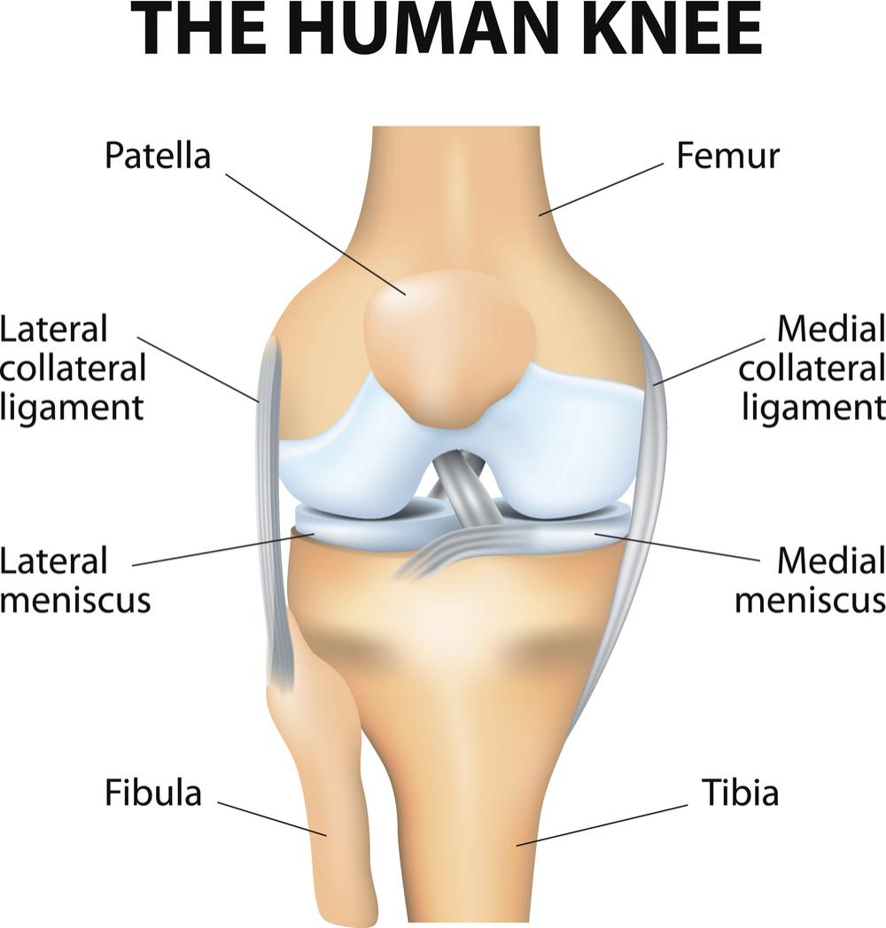
The Knee Anatomy Injuries Treatment And Rehabilitation from cdn-prod.medicalnewstoday.com The hand incorporates countless muscles, bones, tendons and ligaments into simple motion and this chart covers them all. Attaches the calf muscles to the calcaneus, most important muscles for running, jumping, walking etc. Lower back muscle diagram anatomy does degenerative disc disease affect the lower back muscle? The tendon travels along the inside of the forearm on the side of the small finger and crosses the wrist. There are a whole range of structures e.g. Lumbar sprain occurs when ligaments are overstretched or torn. More images for tendon diagram » The fcu tendon is one of two tendons that bend the wrist.
It attaches to the wrist bone, the pisiform, and as well as the 5th hand bone.
Bones, muscles, tendons and nerves which will each give slightly different foot pain symptoms. Attaches the calf muscles to the calcaneus, most important muscles for running, jumping, walking etc. There are a whole range of structures e.g. May 11, 2021 · muscles of lower back diagram in this image, you will find an occipital bone, sternocleidomastoid, trapezius, deltoid in muscles of the lower back diagram. Jul 12, 2020 · muscle anatomy for gym 12 photos of the muscle anatomy for gym muscle anatomy and fitness, muscle anatomy for fitness, muscle anatomy for gym, human muscles, muscle anatomy and fitness, muscle anatomy for fitness, muscle anatomy for gym. The hand incorporates countless muscles, bones, tendons and ligaments into simple motion and this chart covers them all. The achilles tendon is a tough band of fibrous tissue that connects the calf muscles to the heel bone (calcaneus). What is the strongest tendon in the body? What are the major tendons in the body? Tendon, tissue that attaches a muscle to other body parts, usually bones. Intermediate back muscles and c. What are some examples of tendons? A foot pain diagram is a great tool to help you work out what is causing your ankle and foot pain.


0 Komentar